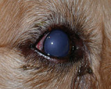
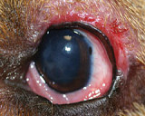
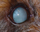
Glaucoma
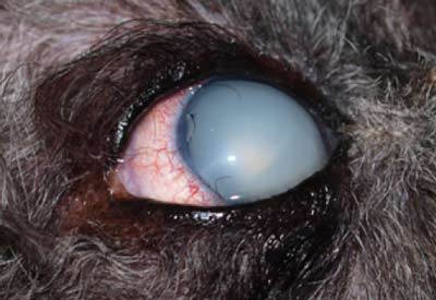
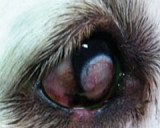
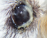
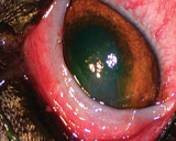
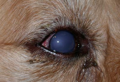 Glaucoma is caused by Increased pressure within the eye. The increased pressure can rapidly cause blindness, and also pain.
It is hard to assess pain in animals, but we find that nearly all our patients are brighter, more active after having glaucoma relieved. Humans described the pain like having a bad headache; you can function, but not happily.
The normal pressure within the eye is 10 to 25 mm Hg. When the pressure is increased (usually > 30 mm Hg) a diagnosis of glaucoma is made. We measure the pressure in the eye (intraocular pressure - IOP) with a tonometer.
Once the eye becomes enlarged the eye is blind.
Causes
Causes for DOGS
Primary Glaucoma in some dogs the outflow of fluid from the eye is blocked by an abnormal drainage angle. Breeds that are affected by this include Basset Hounds, Cocker Spaniels, Australian Cattle Dogs, Fox Terriers, Poodles, Maltese Terriers and Golden Retrievers. Secondary Glaucoma is glaucoma that develops due to inflammation of the eye (uveitis), lens luxation, blood in the eye (hyphaema), or due to growths inside the eye.
Causes for CATS
Glaucoma is usually secondary to long standing inflammation in the eye, or to growths within the eye. We usually try intensive cortisone therapy and if the pressure does not decrease then eye removal or an intrascleral prosthesis is usually indicated.
Management
1. The usual presentation is a dog with a blind, painful eye. We need to treat this eye and make it comfortable.
2. Treatment is given to the good eye to delay glaucoma developing if it is predisposed to glaucoma.
3. This condition may get worse, even with the correct treatment. Drops and tablets seem to work extremely well in humans, but are rarely effective in dogs.
4. Surgery is usually required to control glaucoma, in some cases we can use drops in to the eye once daily. With time we find that some glaucoma cases are not as well controlled with the drops, and surgery may then be required. The management of glaucoma depends on whether or not the eye is still visual or has a chance to regain vision.
Potentially visual eyes
To save vision we use a state of the art Diode Laser to destroy some of the fluid producing cells inside the eye. This is what is commonly done in humans that no longer respond to medications. We generally monitor the patients intraocular pressure frequently over the next few days depending on the pressure readings..
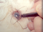
For blind eyes it is important to treat the glaucoma to try and reduce the pain inside the eye. For blind, enlarged eyes we recommend an Intrascleral Prosthesis. For blind eyes that are not enlarged we can laser to control the glaucoma.
Prevention
We can check to see if an eye is predisposed to glaucoma by examining the drainage angle with a special contact lens. This is called Gonioscopy. If the drainage angle is malformed then there is a greater risk of glaucoma developing in the eye. If an eye is predisposed to glaucoma, then lifetime treatment with drops is required. However, even with diligent use of drops it is possible for the eye to develop glaucoma. Prompt recognition and treatment of the glaucoma are essential to save vision.
The early signs of glaucoma
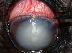
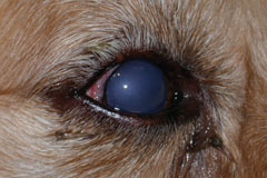
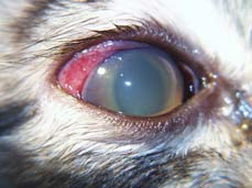
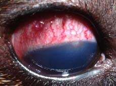
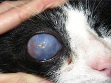
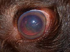
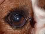
1. Redness of the white of the eye
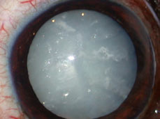
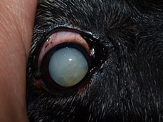
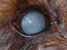
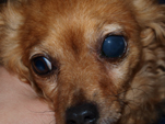
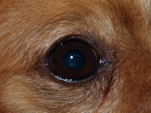
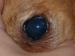
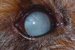
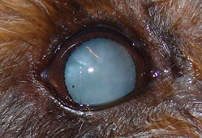
2. Blue, hazy eye
3. Dilated pupil that will not become smaller when a bright light e.g. penlight, is shone into the eye.
 Cataract surgery is a very involved procedure, and there is a lot of preparation prior to and after surgery. The following options will inform you more about cataracts and the procedure, however if you have any further questions prior to making a possible appointment please do not hestitate to call the clinic.