Cloudy eyes in many dogs are due to cataracts and can have a big impact on vision and quality of life. Here at Animal Eye Care we recognise the importance of the human-animal bond and the desire for many owners to help their precious companion animals.
In many cases cataract surgery is an option to restore or improve vision. Surgery is complicated and at Animal Eye Care is performed only by a veterinary eye specialist with use of specialised equipment and an operating microscope. The nursing team are highly experienced in veterinary ophthalmology surgery and expert at monitoring your pet during and after surgery.
The first step in the process is to book an appointment for a consultation with a veterinary ophthalmologist to examine your pet’s eyes. A thorough examination is performed of the entire eye to determine whether your pet is a suitable candidate for cataract surgery.
If cataract surgery is an option we will dispense some eye drops to prepare the eyes for surgery. We will also liaise with your regular veterinarian about your pet’s general health, dental health and whether they are sufficiently healthy for a general anaesthetic.
The success rate of cataract surgery is around 90-95% in most cases. The caring team at Animal Eye Care take great delight in restoring the vision of blind animals and several of our nurses say that it is the favourite part of their job!
Diabetic dogs are at greatly increased risk of developing cataracts. Here at Animal Eye Care we have dedicated procedures and protocols for dealing with your pets special health needs.
To make an appointment for examination of your pet’s cloudy eyes please contact Animal Eye Care on (03) 9563 6488 to arrange a suitable time and location.
CATARACTS AND CATARACT SURGERY IN MORE DETAIL
What is a cataract?
A cataract is an opacity of the lens. The lens is normally clear and transparent. When a cataract forms the lens itself goes cloudy. A cataract is not a film or coating of the eye, but the inside of the lens – the lens protein goes cloudy. A cataract can affect just a small part of the lens, or it can affect the entire lens. Small cataracts may not have any affect on vision. If the cataract involves the whole lens in both eyes then vision will be affected. In more severe cataracts you may notice that the pupil, which normally appears black, has undergone a colour change and becomes bluish or white.
What can cause a cataract?
In most cases we do not find a cause for the cataracts, they just develop. The most common causes we find include diabetes, inherited causes, and PRA (progressive retinal atrophy). Breeds seen at Animal Eye Care with inherited cataracts include Cocker Spaniels, Poodles, Australian Cattle Dogs, Maltese, Miniature Schnauzers, Labradors, Boston Terriers, Bichon Frise and Golden Retrievers. It is difficult (except in some diabetics) to look at a cataract and determine the cause.
What should I do if I suspect my pet has a cataract?
If you or your local veterinarian has noticed clouding of the eyes, or poor vision, then your pet should be examined by a veterinary ophthalmologist and given a comprehensive eye examination. This will involve dilating the pupils and examining the lens with special equipment. The nature of the cloudiness will be determined. If a cataract is present then its size, position and type will be determined. It is best to perform this examination before the cataracts become too developed.
Why do we need to check the retina the nerve tissue?
The retina (the nerve tissue at the back of the eye) must be healthy for cataract surgery to restore vision. Many purebred dogs (and their crosses) such as Labradors, Australian Cattle Dogs, Australian and Silky Terriers, Poodles and Cocker Spaniels can have retinal disease. The most common retinal disease is PRA (progressive retinal atrophy) which is an inherited disease. PRA in the early stages can cause poor night vision, then later poor day vision but also often causes cataracts to form. In many cases the early signs of PRA are missed, even by the most diligent of owners, and it is assumed that the cataracts are the cause of the vision loss. In these cases the dog is blind because of the cataracts and the PRA, and cataract surgery will not restore vision.
Electroretinography (ERG)
Electroretinography (ERG) If we cannot see the retina because the cataract is too cloudy, an ERG (electroretinogram) will be performed. This is an electrical test of the retina. This will require half a day in hospital and sedation. Using a computer we measure the electrical response to bright lights flashed into the eye.
If the retina is healthy the ERG will show a normal wave pattern. If the retina is diseased with PRA the ERG is flat. The ERG will not show early retinal detachments. PRA is diagnosed with the ERG when we are not able to see through to the retina because the cataracts are too cloudy. If the PRA is only in the early stages (as detected with the ERG) then cataract surgery may improve vision for 6 to 18 months, but as the PRA is progressive the patient will eventually go blind. The ERG is a test of the electrical function of the eye. 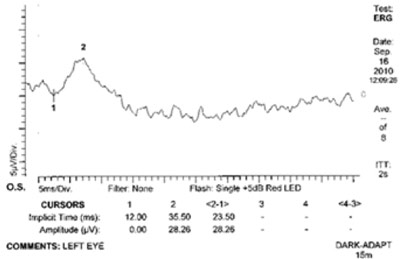
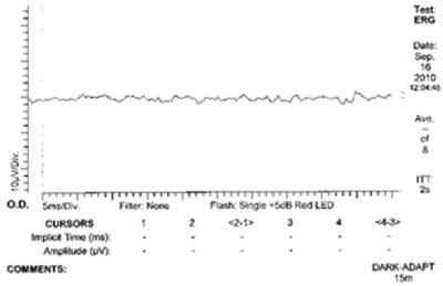
[L-R] ERG: Good Response & ERG: Poor Response.
Ultrasonography
As part of the general workup before surgery we may perform ocular ultrasonography. This is often done with the ERG. The ultrasound is used to image the inside of the eye; in these cases we are checking to make sure that the eye is healthy enough for surgery. In particular we are looking for advanced degenerations of the vitreous, and or retinal detachment. The ultrasound can be considered as a structural examination of the eye.
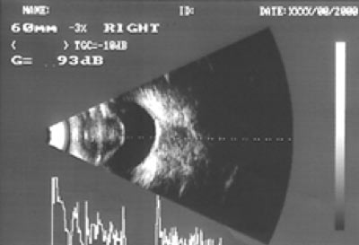
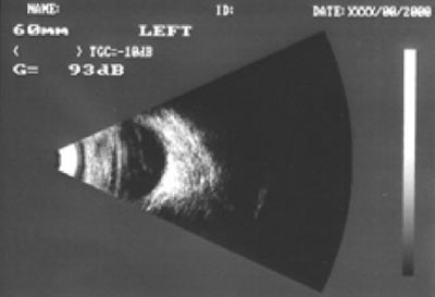
[L-R] Ultrasound: Healthy Retina & Ultrasound: Retinal Detachment.
What is cataract/lens induced uveitis (inflammation)?
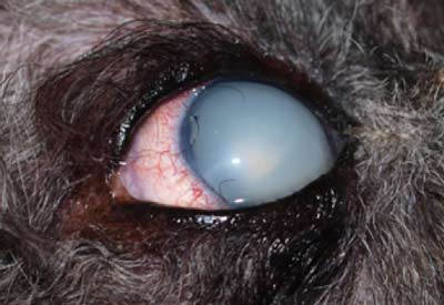 As cataracts form, the lens protein inside the eye changes. This change may allow the lens protein to leak into the inside of the eye. This causes inflammation inside the eye, and this must be treated. Lens induced uveitis can cause painful outcomes such as glaucoma, and or lens luxation. Pre-operative lens induced uveitis will lower the success rate of cataract surgery.
Lens induced uveitis can be controlled in most cases with long-term drops and in some cases tablets. Surgery is often helpful in some cases to help control the lens induced uveitis. The cataracts that develop because of the PRA will often cause this lens-induced uveitis. Watch for signs of redness on the whites of the eye. If treated early, painful outcomes such as glaucoma and lens luxation can be avoided.
What is the treatment for cataracts?
Surgery is the only effective method to remove the cloudy lens from the eye. There are no known preventative treatments for cataracts. From time to time various drops, lotions, pills, and special diets have been suggested to help with cataracts. These have not been proven to help dissolve or reduce a cataract. More details on cataract surgery are outlined on our Cataract Surgery handout.
Should my pet have cataract surgery?
Most blind animals cope very well as they have good senses of smell and hearing, and they know their home environments extremely well. Successful cataract surgery can greatly improve an animal’s quality of life. Factors that need to be considered include the patient’s age, general health, and the health of the eyes themselves. In some older dogs we often ask your local veterinarian to run blood and urine tests before surgery. The decision on whether to remove a cataract is a joint one between you, your local veterinarian and Animal Eye Care. Please feel free to speak to us at any time if you have any additional questions regarding cataracts or cataract surgery.
CATARACTS SURGERY POST-OPERATIVE CARE IN MORE DETAIL
Cataract surgery is a complicated procedure that takes place inside the eye. The post-operative instructions are strict and should be adhered to for best results.
Post-operative checks:
A minimum of 4 visits are required over the first 4-6 weeks after surgery. Usually these are the day after surgery, around a week after, two weeks after and a month after. We may recommend extra visits if we are concerned about complications.
The first six weeks of visits are included in the surgery fee. There is no charge if we see your pet at East Malvern during regular clinic hours Monday to Friday. Post-op checks seen at out-clinics (Point Cook, Hoppers Crossing, Essendon, Frankston, Corio) will incur a charge of $45. Post-ops seen at rural clinics (Albury, Moe, Warrnambool, Bendigo) will incur a charge or $60. Post-op checks seen out of regular hours at East Malvern will incur an emergency fee ($200 plus any diagnostic tests and medications).
After the first six weeks we then recommend a revisit at three months after surgery and then 6-12 monthly to ensure the health of the eye. There is a charge for these consultations.
Medications after surgery: Eye drops to control infection, pressure and inflammation are given immediately after surgery. Some of these may be required long term. Anti-inflammatory oral medications are given for six weeks.
Activity after surgery: Strict rest is required for the first 7-10 days after surgery. It is important to
- Keep the E collar on for 7 days
- Try to stop or minimise barking
- Stop ball games or toys that involve shaking the head (for six weeks after surgery!)
- Lead or harness walking for toileting only
Gentle lead walking along quiet streets is allowed after 7-10 days.
Off-lead exercise is allowed after 4-6 weeks.
Bathing is allowed two weeks after surgery.
Contact us if you see any of the following:
- Poor vision
- Squinting
- Watery or mucky discharge
- Redness
- Cloudiness or a blue haze to the eye
We will show you how to perform a menace test to check vision and how to use a bright light to check normal response by the pupils.
If you are at all concerned we would like you to contact us promptly. During clinic hours you can call 03 9563 6488
Outside of clinic hours you can call 03 9572 1966 (there is a vet on call until 10 pm weekdays or 6 pm weekends). Please leave a message outside of these hours or see an emergency veterinarian if very concerned.
