Notes on surgical technique, Animal Eye Care
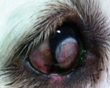
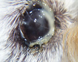
Modification of the Rhea Morgan Pocket Technique for replacement of prolapsed third eyelid glands (Cherry Eye).
In this procedure, an elliptical incision is made in the superficial conjunctiva overlying the third eyelid gland. This is excised and the site closed with a continuous 6/0 absorbable suture taking care to bury the knots.
Notes on surgical technique:
- Pemedication with carprofen to reduce postoperative swelling.
- Use magnification e.g. Voroscope, Welch Allyn Lumiview
- Prep the area with 1% betadine solution. Place on Opsite® drape across the eye then make an incision from medial to lateral canthus. Insert an eyelid speculum (eyelid retractor).
- Elevate and expose the bulbar surface of the third eyelid and its gland using mosquito forceps by placing one at each end of the gland.
- Make an elliptical incision of the conjunctiva overlying the gland. See diagram 1. Join the ends of the incisions. The proximal incision should be over 5mm from the TE margin. An incision too close will result in prominence of the gland. The ellipse should be 8-12mm wide, depending on the size of the gland and breed of dog.
- Dissect off the conjunctiva. Take care to keep the plane of dissection very superficial i.e. just remove the thin conjunctiva.
- Check the third eyelid cartilage – if everted (kinked) dissect superficially to remove a portion to allow the TE to sit flat once the surgery site is closed. This can be difficult and good magnification is essential.
- For enlarged glands, place 2 or 3 buried simple interrupted sutures to reduce tension on the continuous sutures. See diagram 1. This will make the gland sit into the pocket. Use 6/0 Polysorb® or vicryl or dexon i.e. absorbable multifilament.
- Suture the ellipse closed. Bury the knot at the start. We use a Connell-type suture pattern i.e. continuous horizontal mattress suture pattern that inverts the wound edge. See diagram 2. After a couple of bites, the gland should be able to be pushed down into the pocket (if tension sutures haven’t previously been placed) making the remainder of the sutures easy to place.
- Bury the knot again at the completion of closure. We often finish the closure then pass the suture through to the outer side of the third eyelid and take a small bite then tie the knot here. See diagram 3. This way there is no chance of suture irritation from a knot.
- Remove the forceps. The TE should sit normally, however if the gland if very enlarged the edge of the TE may sit slightly away from the cornea. The TE may sit across the eye for a few days.
- Send home on Tricin® ointment TID for 7-10 days. Recheck at 7-10 days post operatively. Do not attempt surgery in a Bulldog, Neopolitan Mastiff or other Mastiff breed unless you are highly experienced. These cases should be referred.
