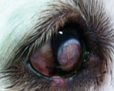
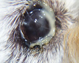
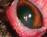
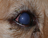
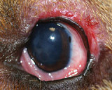
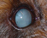
Differential Diagnosis include: 1. Haemangioma (usually looks like a blood blister) 2. Melanoma (usually black but can be amelanotic) 3. Limbal melanocytoma (smooth black swelling arising at the limbus) 4. NGEK Nodular granulomatous episcleriokeratoconjunctivitis (pale pink to cream-coloured swellings)
Conjunctival Haemangioma/Haemangiosarcoma
 Conjunctival Haemangioma/Haemangiosarcoma - These tumours are seen most commonly in middle-aged Kelpies, Border Collies and Aust. Cattle Dogs. They are thought to be secondary to UV exposure and are usually situated in the conjunctiva adjacent to the lateral limbus. In the earliest form it may appear as swollen blood vessels and hyperaemia and may respond to topical cortisone drops. Once they are blood-blister-like in appearance, surgery is required. Excision of the mass with 3-5mm margins and cryotherapy is usually curative.
They do not appear to be related to haemangiosarcoma seen elsewhere in the body. Malignancy at pathology (i.e. sarcoma vs haemangioma) is not associated with poorer treatment outcomes.
In all cases of conjunctival mass surgery, the cut edges of the conjunctiva must be sutured to the sclera. Cortisone tablets are used post-operatively to treat possible uveitis associated with cryotherapy and the eye is checked at 1-2 weeks. Owners are advised to monitor the eye daily for signs of uveitis. Cortisone drops are used once healing is complete to reduce hyperaemia/scar tissue.
Conjunctival Melanoma
Conjunctival Melanoma: Melanomas may arise from any pigmented naevus of the conjunctiva – eyelid, bulbar or third eyelid. They are usually raised, soft, black masses. Treatment involves excision with 5mm margins (when possible) and cryotherapy of the base and surrounding tissue in a similar method to that described above. Cryotherapy is usually more extensive compared to haemangiomas due to size and may result in uveitis; appropriate uveitis treatment is commenced in case. Extensive tumours may not be treatable and eye removal may be required.
Limbal Melanocytoma
 Limbal Melanocytoma: These tumours arise from limbal melanocytes and appear as smooth, usually round black swellings at the limbus. They extend into the cornea and back into the sclera for variable distances. Limbal melanocytomas are usually benign but can continue to grow and interfere with function of the eye.
Limbal Melanocytoma Treatment is surgical excision via keratectomy and sclerectomy. It is possible to completely resect the pigmented tissue in some cases. Adjunctive treatment of the base with cryotherapy, diode laser or Strontium 90 is necessary. If the growth is full thickness, the structure of the globe is maintained via autogenous grafting with third eyelid cartilage.
Nodular Granulomatous Episcleriokeratitis

Nodular Granulomatous Episcleriokeratitis: NGEK appear as benign swellings of the sclera with associated conjunctival hyperaemia. The most common anatomic position is lateral and ventrolateral sclera adjacent to the limbus, but any area may be affected. Breeds more commonly affected include Rottweilers and Shelties.
Nodular Granulomatous Episcleriokeratitis Treatment is the use of topical cortisone drops and systemic cortisone therapy. Most cases respond well however refractory cases may respond to oral tetracycline (500mg TID) or doxycycline (5mg/kg BID to TID) and nicotinamide (250-500mg TID). Cases refractory to this treatment may respond to cryotherapy or Strontium 90 brachytherapy. Recurrence is likely and long term therapy is usually required; dose and frequency rates may reduced to minimal levels to prevent recurrence and increased in response to flare-ups.
In all cases, clients must be warned to monitor for recurrence as early treatment will result in more complete excision and favourable outcome.
